Out of mind, out of sight: The blind man who can 'see' obstacles
Experiments on a blind man who can ‘see’ to avoid obstacles could have huge implications for the visually impaired

In a darkened room, a blind man walks along a white line in the shape of a large ellipse.
He is taking part in an experiment which I have been invited to watch, at the University of Geneva in Switzerland. At one point, the scientist running the experiment, Beatrice de Gelder, asks me to stand on the white line, in the man’s path. I mustn’t move or make a sound. When he is about a metre away, he comes to a halt and asks: “Is somebody there?”
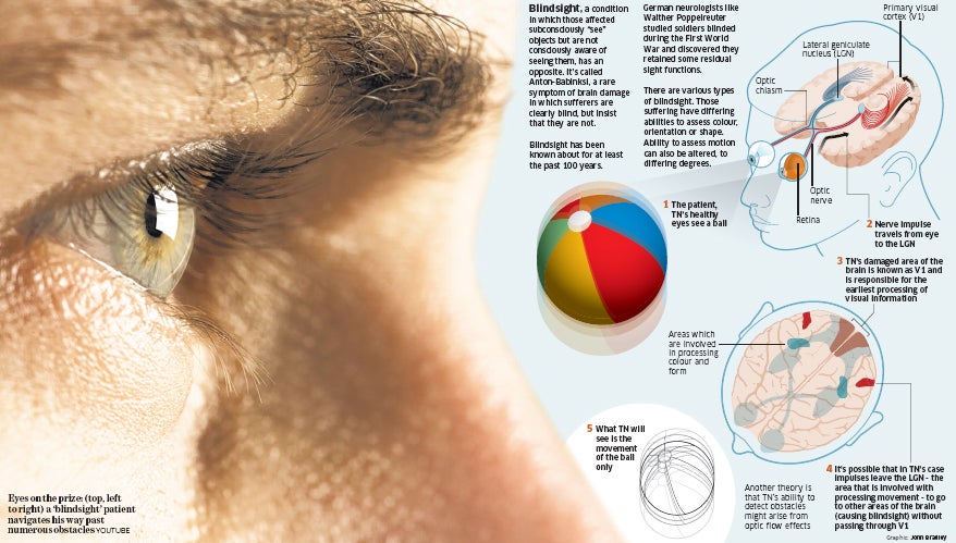
TN, as the blind man is known, suffered a stroke in 2003 which destroyed an area at the back of his brain that processes visual information: the primary visual cortex. The stroke affected only one hemisphere of his brain. What places TN in a category of his own, at least as far as the annals of science are concerned, is that about a month later he suffered a second stroke which wiped out the primary visual cortex on the other side of his brain. Suddenly, though his eyes were healthy, he became blind.
TN’s blindness is unusual, however, because he can still see in some situations, although he is unaware that he does so – a phenomenon known as blindsight. The most striking demonstration of this came two years ago, when de Gelder, a neuropsychologist at Tilburg University in the Netherlands and Harvard Medical School in the US, and neuropsychologist Alan Pegna of Geneva University and others, asked him to walk down a corridor which they had arranged like an obstacle course, having littered it with tripods, filing trays and boxes. He navigated his way successfully through the obstacles, though he said he saw none of them.
It’s well-known that hearing can become more acute in blind people, so to exclude the possibility that TN was relying on sound cues to negotiate his way through the obstacles the researchers asked him to repeat the experiment wearing earplugs. His performance didn’t change. “So it’s not auditory information that’s helping him,” says Pegna. “On the other hand, when we blindfolded him, he started bumping into the obstacles.”
Pegna first tested TN in 2004, and at that time he identified another visual skill that had been partially preserved in him: the ability to recognise facial expressions of emotion. When TN was asked to look at a series of angry, happy and fearful faces, and to guess what expression was being displayed, he guessed correctly more often than he would have had he been responding at random.
The effect wasn’t as dramatic as in some blindsight patients whose primary visual cortex has been damaged on one side of the brain only, and to whom faces have been presented in the blind half of their visual fields – some of whom responded correctly 90 per cent of the time. TN’s success rate was closer to 60 per cent. Nevertheless, he seemed to have retained some ability to detect emotional expressions without being consciously aware of them. And when the researchers put him in a brain scanner and watched how his brain responded, they found that one of his amygdalae – a pair of brain structures known to be sensitive to emotion – became significantly more active when he was looking at emotional expressions than when he was looking at neutral ones.
Pegna and de Gelder would like to know what brain pathways TN is using to see. The mechanics of vision are complex and still not well-understood, but scientists know that the main fibre tract leaving the retina at the back of the eye passes via a structure called the lateral geniculate nucleus (LGN) to reach the primary visual cortex, or V1 – the area that is damaged in TN. V1 is responsible for the earliest and most basic processing of visual information. It essentially parses the visual scene according to colour, form and motion, sending the results to other visual areas of the brain’s cortex for more higher-order processing.
TN’s case raises two possibilities. The first is that fibres leaving the LGN don’t just project to V1, but that some go directly to the other visual areas without first passing through V1. There is now good evidence, for example, that the LGN has a direct connection to an area called V5, which is involved in processing movement, and which is probably intact in TN.
In 1993, visual scientist John Barbur of London’s City University, and colleagues, used brain imaging to show that V5 still responded to input from the retina in a patient with damage to V1 who was able to detect moving visual stimuli. And this year, researchers at the US National Institute of Mental Health in Bethesda, Maryland, led by Michael Schmid, showed a similar effect in V1-damaged macaque monkeys. Schmid’s team also showed that when the LGN was damaged in addition to V1, the V5 response was abolished, and the monkeys could not detect visual stimuli in tests.
De Gelder suspects that TN’s ability to detect obstacles might arise from optic flow effects as he moves towards them. It’s a hypothesis she has yet to prove, but it is telling, she says, that his performance on the obstacle course deteriorates when he wears goggles that block his peripheral vision. Optic flow is exemplified by the opening sequence of the film Star Trek, in which stars moving past the starship Enterprise give viewers the impression they are moving into the screen, and the effect is most-pronounced at the periphery of the visual field.
The other possibility is that TN is making use of a separate visual pathway altogether. This second pathway bypasses the LGN and is mediated instead by a structure in the brainstem called the superior colliculus. The brainstem is the oldest part of the brain, evolutionarily speaking, and this pathway is not accessible to consciousness. This month, TN was back in Geneva for more tests. Though the scientists haven’t yet found any hard evidence of regeneration or reorganisation in his brain since his strokes, his condition seems to have improved. Larry Weiskrantz, an emeritus professor of psychology at Oxford University, has identified two subtypes of blindsight: type 1, in which patients perceive nothing at all, and type 2, in which they have an impression of seeing something, but cannot say what it is.
“TN seems to have shifted from type 1 to type 2,” says Pegna. He now complains of visual hallucinations, for example. De Gelder says that in healthy people, the streams of information that makeupvision – the perception of colour, motion and form – are integrated into one seamless visual percept, and that this integration may be performed with the help of consciousness. Because TN is not conscious of what he sees, he may have lost that coherent quality of his perceptual experience, while retaining some or all of the separate components of it.
Last month, when I had the opportunity to watch him, TN negotiated obstacle courses more efficiently than he did two years ago, though he still wasn’t comfortable relying on his subconscious skills, asking for someone’s arm when he left the testing situation. And he remained unable to identify the obstacles. When, in de Gelder’s experiment that had him follow a white line on the floor, I replaced a wooden stick as the obstacle, he still asked: “Is someone there?”
When it came to detecting facial emotion, his slight recovery seemed, counter-intuitively, to have impaired his performance. Though his amygdalae responded as before in brain scans, his rate of successful identification of the facial expressions he was looking at was now no higher than 50 per cent, which is the same as getting it right by chance. Pegna thinks he may have been trying to interpret those vague impressions that he has. In other words, conscious processes may have been interfering with his subconscious seeing.
Ultimately, the researchers’ goal is to understand how the normal brain sees. The subconscious visual pathway is present in all of us, for example, but do healthy people use it? Is it normally suppressed by the conscious pathway, or does it constantly feed into and influence our conscious visual experience, perhaps by directing our attention to, and so amplifying the signal from, relevant aspects of the visual scene? For the moment these questions have no answers, but with the continued cooperation of TN and other visually impaired patients, Pegna and de Gelder hope it won’t be long before they do.
Subscribe to Independent Premium to bookmark this article
Want to bookmark your favourite articles and stories to read or reference later? Start your Independent Premium subscription today.

Join our commenting forum
Join thought-provoking conversations, follow other Independent readers and see their replies