Brain tumour survival could be improved by drink which makes cancer cells glow pink in surgery, new study suggests
'Desperately needed' treatment could help surgeons identify cancerous tissue
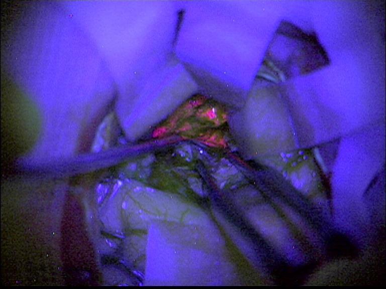
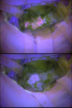
A drink which makes cancer cells glow pink under fluorescent light could help surgeons to identify and remove aggressive brain tumours and minimise the damage to healthy brain tissue, an NHS trial has suggested.
Before undergoing surgery, patients were urged to drink the chemical substance before surgery which accumulates in cells of aggressive, fast-growing glioma, the most common group of brain tumour, by researchers led by Birmingham University.
Operating surgeons have to remove as much of the cancerous tissue as possible to ensure they do not regrow into tumours and limit the need for aggressive follow-up chemo or radiotherapy. Fast growing (high grade) tumours are particularly important to identify as they are most likely to cause recurrence.
“The advantage of this technique is that it may highlight more quickly high-grade disease within a tumour during neurosurgery. What this means is that more of the tumour can be removed more safely and with fewer complications, and that’s better for the patient," said Professor Colin Watts, neurosurgeon with the University of Birmingham who led the research.
“These results show that the marker is very good at indicating the presence and location of high-grade cancer cells."
It is thought the liquid could also help surgeons consider treatment next steps while operating, without having to wait for pathology results to confirm whether tumours are high or low grade.
The preliminary study only measured whether the chemical – known as 5-aminolevulinic acid (5-ALA) worked and did not test patient survival after surgery.
Doctors saw the marker in 85 out of 99 patients and later tests confirmed that 81 of these had high grade tumours, one had slow growing tumours and three could not be identified.
There were no fast growing tumours identified where the marker did not work, although some tumours could not be identified in pathology tests.
The researchers said in many cases survival is a matter of months not years – and this treatment has the potential to improve this.
"There is a desperate need for better treatments for brain tumours and to achieve that we need more high quality research in this area,” said Professor Anthony Chalmers, a clinical oncologist at Glasgow University who was not involved with the study.
“This technique provides on-the-spot information to help surgeons tailor the operation according to the location, size and grade of the tumour.
“We know that patients who have near total removal of their tumour have better outcomes, so we are optimistic that, in the long term, these new data will help to increase survival times for glioma patients.”
The results are not yet published but are being presented at the National Cancer Research Institute (NCRI) Conference in Glasgow on Sunday.
Join our commenting forum
Join thought-provoking conversations, follow other Independent readers and see their replies
Comments
Bookmark popover
Removed from bookmarks