Scientists create ‘mini beating heart’ in a petri dish in major medical breakthrough
Groundbreaking development paves the way to develop treatments for a wide range of heart defects
A team of scientists have developed a beating “mini-heart” formed in only a small petri dish – paving the way for future treatments.
Using 35,000 pluripotent stem cells, which are spun into a sphere using a centrifuge – a fast-spinning machine – the groundbreaking development is complete, Jam Press reports.
In a bid to learn more about the earliest stages of the human heart and develop treatments for a range of different defects, experts have carried out the process.
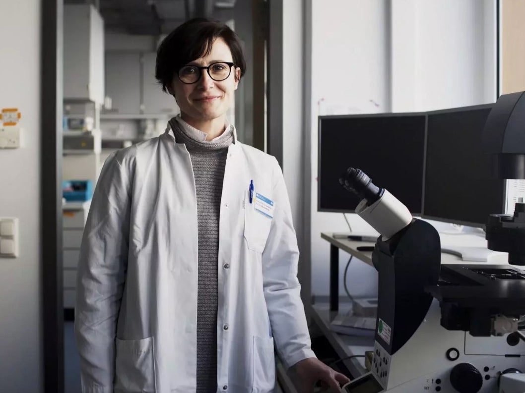
The study was headed by Dr Alessandra Moretti, Professor of Regenerative Medicine in Cardiovascular Disease, at the Technical University of Munich (TUM) in Germany.
Under a set protocol, various signalling molecules were added to the cell culture during a period of several weeks.
Dr Moretti explained: “In this way, we mimic the signalling pathways in the body that control the developmental programme for the heart.”
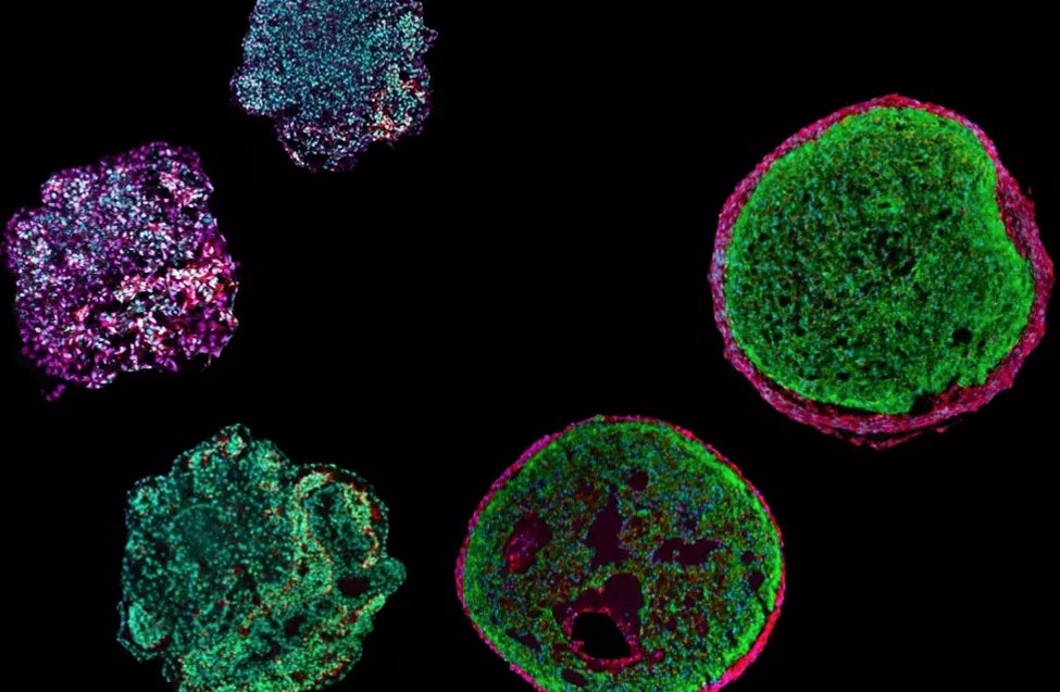
The resulting “organoid” measures about half-a-millimetre in diameter and while it doesn’t pump blood, it can be electrically stimulated and contracts like a human heart.
It’s believed the human heart starts to form around three weeks after conception, when most mothers are still unaware of the pregnancy.
While studies on animal hearts cannot be transferable to human research, experts are hopeful that the groundbreaking development can provide answers.
The TUM team is the first in the world to successfully create an “organoid” with cells of the outer layer of the heart wall (epicardium) and heart muscle cells (cardiomyocytes).
Study author Dr Anna Meier said: “To understand how the heart is formed, epicardium cells are decisive.
“Other cell types in the heart - for example in connecting tissues and blood vessels - are formed from these cells.
“The epicardium also plays a very important role in forming the heart chambers.”
After analysing individual cells, researchers discovered that precursor cells recently found in mice are formed around the seventh day of developing the “organoid.”
The epicardium, the innermost layer of the pericardium, is formed from these cells.
Scientists hope these insights can shed light on why a foetal heart is able to repair itself, something an adult heart is almost entirely incapable of.
With this information, researchers could develop new treatments for a wide range of heart conditions, including heart attacks.
Join our commenting forum
Join thought-provoking conversations, follow other Independent readers and see their replies
Comments
Bookmark popover
Removed from bookmarks