Wellcome Image Awards: The most striking images from the world of science, including breast cancer cells under chemical attack and a photographer’s own kidney stone
Pictures that focus on everything from cells to plant life and embryos are celebrated at the Wellcome Image Awards

Sign up for a full digest of all the best opinions of the week in our Voices Dispatches email
Sign up to our free weekly Voices newsletter
Some of the most striking images from the world of science – from a photographer’s own kidney stone to the X-ray of a bat killed by a cat – will be going on public display following an awards ceremony on Tuesday night that will reveal the winner of the 2014 Wellcome Image Awards.
The winning entry will be chosen from a shortlist of 18 images, including a scanning electron-micrograph of a head louse egg, a 3D computed tomography image of a seal skull and a cross-section of a flower bud – all showing in minute detail the visual wonder of the natural world.
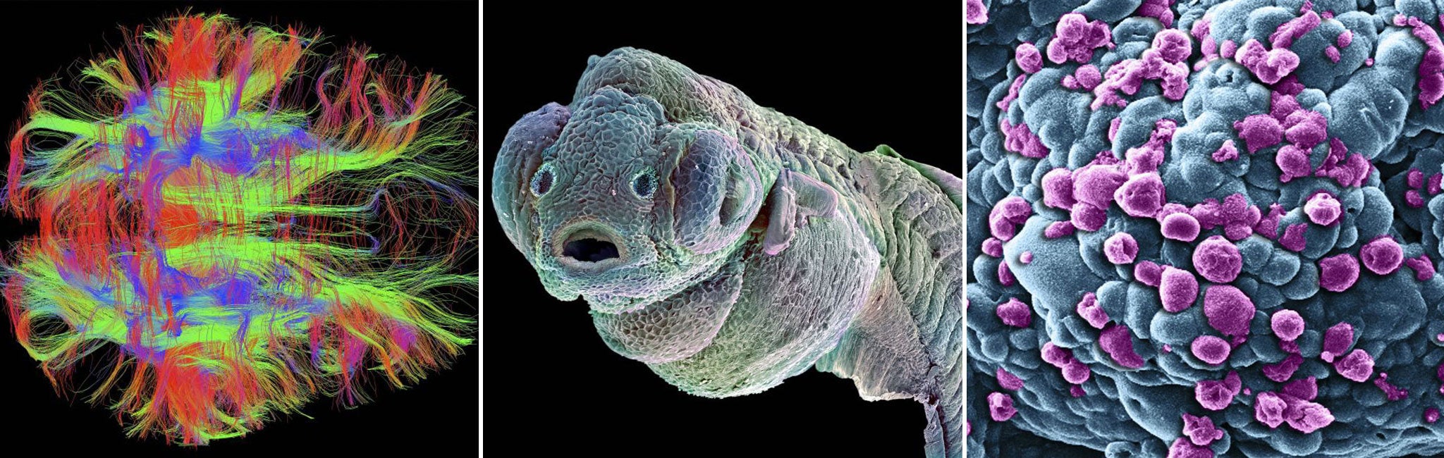
Among those shown here is a bird’s eye view of the nerve fibres in a normal, healthy adult brain (1). The communicating brain cells are visualised by diffusion-weighted magnetic resonance imaging, which is a specialised type of MRI scanner measuring the movement of water in many directions – colour coded here to indicate the direction of travel for the nerve fibres.
The Wellcome Image Awards
Show all 18The image, taken by Dr Zeynep Saygin, of the Massachusetts Institute of Technology, is being used to understand the patterns of connectivity in the brain to better understand the functioning of this most complex of human organs.
“I use connectivity patterns to predict brain function in healthy adults, and see whether the same patterns are present in children. This is especially interesting for mental functions that only emerge with relevant experience, such as reading,” Dr Saygin said.
In another image (5), a scanning electron microscope shows a cluster of breast cancer cells (blue) being attacked by nanometre-sized particles carrying the anti-cancer drug doxorubicin, which is already killing some of the tumour cells (purple) by a process of programmed cell death, or apoptosis.
Doxorubicin does not distinguish between normal cells and cancer cells so it is important to visualise how the nanoparticles are able to target the site of the tumour without causing collateral damage to the healthy tissues of the body.
Subscribe to Independent Premium to bookmark this article
Want to bookmark your favourite articles and stories to read or reference later? Start your Independent Premium subscription today.
Join our commenting forum
Join thought-provoking conversations, follow other Independent readers and see their replies